,,Ug-PhNwkkiQ,UCnXOULz77TMQupryNOK9u_w, Health,Knowledge, channel_UCnXOULz77TMQupryNOK9u_w, video_Ug-PhNwkkiQ,Hello, I'm Dr. Aizaz here with a quick announcement. The lecture notes for the Head and Neck and Pharyngeal arches embryology lectures are now available in your member's area. You can download the PDF notes by going to the member's area, clicking on the lecture notes, and then downloading the PDF. Let me show you how to download them.
,,sZPPSoFBGgA,UCnXOULz77TMQupryNOK9u_w, Health,Knowledge, channel_UCnXOULz77TMQupryNOK9u_w, video_sZPPSoFBGgA,Development of Telencephalon: From Neural Tube to Complex Brain Structures
In this comprehensive embryology lecture, we delve into the intricate development of the telencephalon, a fascinating journey that results in the formation of crucial brain structures including the cerebral cortex, corpus striatum, and hippocampus. This video serves as an essential resource for medical students, USMLE aspirants, and healthcare professionals, offering both fundamental concepts and advanced developmental processes.
Our journey begins with the basic development of the telencephalon, where we explore how the initial telencephalon impar gives rise to bilateral evaginations that form the lateral ventricles. We carefully examine the formation of the lamina terminalis and the development of cerebral hemispheres, including the crucial establishment of the interventricular foramen of Monroe. Through detailed 3D animations, we demonstrate how these early structures set the foundation for more complex brain development.
The lecture then progresses to an in-depth exploration of the ganglionic eminences and their derivatives. We examine the medial, lateral, and caudal ganglionic eminences, tracking the intricate migration patterns of neuroblasts as they form various brain structures. The video clearly illustrates how these migrations lead to the formation of the corpus striatum components and inhibitory interneurons. Special attention is given to the concept of pallium and subpallium, providing a clear framework for understanding these developmental processes.
A significant portion of the lecture focuses on cerebral cortex development, where we witness the remarkable formation of the major lobes - frontal, parietal, temporal, and occipital. Through advanced visualization techniques, we demonstrate the development of the insula and opercula, and the progressive remodeling of cortical structures. The formation of sulci and gyri is explained in detail, along with the development of olfactory structures, providing a comprehensive understanding of how the brain achieves its characteristic appearance.
The video then takes you through the fascinating development of subcortical nuclei, including the formation of the corpus striatum and its components. We explore how the caudate nucleus achieves its characteristic C-shape, and the development of the putamen and globus pallidus. The role of the internal capsule in striatum formation is clearly demonstrated, along with the development of the amygdala, using state-of-the-art animations to illustrate these complex processes.
Particular attention is given to hippocampal formation, where we examine its development from the medial pallium. The intricate processes leading to the formation of the hippocampal cortex, dentate gyrus, and indusium griseum are carefully explained. We also explore the development of the parahippocampal gyrus and demonstrate why these structures collectively resemble a seahorse, leading to their namesake.
The lecture concludes with a comprehensive overview of commissural development, including the formation of the corpus callosum, fornix, and hippocampal commissure. The integration of these commissural systems with other brain structures is clearly illustrated, providing a complete picture of brain connectivity development.
Throughout the video, we employ advanced visualization techniques and clear diagrams to make these complex developmental processes more accessible and understandable. The content bridges the gap between basic embryology and advanced neuroanatomy, making it an invaluable resource for understanding both normal brain development and related congenital anomalies.
Whether you're preparing for medical examinations or seeking to deepen your understanding of brain development, this video offers valuable insights into one of the most complex aspects of human embryology. Visit medicovisual.com for additional resources and study materials, and subscribe to our channel for more detailed medical lectures and visual guides.
#Embryology #Neuroscience #MedicalEducation #USMLE #BrainDevelopment #MedicalAnimation #Telencephalon #Neuroanatomy #MedicalStudent #CerebralCortex
,development of pituitary gland,hypophyseal gland,Pituitary gland development,hypophysis,adenohypophysis,neurohypophysis,Rathke's pouch,stomodeum,oropharyngeal membrane,embryology,pars distalis,pars nervosa,pars tuberalis,pars intermedia,craniopharyngioma,optic chiasma,endocrinology,neuroembryology,diencephalon,hypothalamus,prosencephalon,Q1c_XpKyymQ,UCnXOULz77TMQupryNOK9u_w, Health,Knowledge, channel_UCnXOULz77TMQupryNOK9u_w, video_Q1c_XpKyymQ,In this visual lecture Dr. Aizaz from Medicovisual.com provides an in-depth exploration of the intricate process of pituitary gland development during embryogenesis. Building upon previous discussions on the development of the hypothalamus as a derivative of the diencephalon, this lecture delves into the dual embryological origins of the pituitary gland, its structural components, and their functional significance.
The lecture begins by situating the pituitary gland anatomically beneath the hypothalamus. Dr. Aizaz explains that the pituitary gland, also known as the hypophyseal gland, derives its name from "hypo" meaning "under" and "physis" meaning "growth," highlighting its position as an undergrowth of the hypothalamus. This gland appears as a scrotum-shaped structure at the ventral part of the hypothalamus, though humorously noting it has no relation to the scrotum beyond its shape.
Emphasizing the gland's composition, Dr. Aizaz outlines that the human pituitary gland consists of two main parts:
1. Anterior Pituitary (Anterior Lobe): Also called the adenohypophysis or pars distalis, this is the true glandular component responsible for the synthesis, storage, and release of hormones. Derived from epithelial tissue, it plays a crucial role in endocrine function.
2. Posterior Pituitary (Posterior Lobe): Known as the neurohypophysis or pars nervosa, this is not a true gland but rather an extension of the hypothalamus. It stores and releases hormones produced by the hypothalamus but does not synthesize hormones itself.
The lecture delves into the embryological development of these two parts:
1. Development of the Posterior Pituitary:
The posterior pituitary originates from a downward extension of the hypothalamus. Specifically, an outgrowth from the ventral part of the hypothalamus forms the posterior pituitary. This extension, called the infundibulum, develops into the pituitary stalk and the pars nervosa. Since it is an extension of neural tissue, it is termed the neurohypophysis.
2. Development of the Anterior Pituitary:
The anterior pituitary arises from an upward invagination of the ectodermal tissue from the stomodeum, which is the primitive mouth or oral cavity in the embryo.
This invagination forms a pouch known as Rathke's pouch, named after the scientist who first described it.
Rathke's pouch grows upward toward the neurohypophysis, eventually pinching off from the stomodeum and differentiating into the anterior pituitary. Being derived from ectodermal tissue, the anterior pituitary is an epithelial glandular structure, hence the term adenohypophysis.
Dr. Aizaz provides a detailed explanation of the stomodeum and the oropharyngeal (buccopharyngeal) membrane:
The oropharyngeal membrane is a bilayered structure composed of ectoderm and endoderm with little to no mesoderm between them.
The stomodeum is the ectodermal depression anterior to the oropharyngeal membrane and plays a pivotal role in forming the anterior pituitary.
He clarifies common misconceptions between these two terms, emphasizing that while they are related, they are distinct structures.
Visual 3D animations illustrate the formation and transformation of Rathke's pouch:
Rathke's pouch, initially connected to the stomodeum, eventually loses this connection as it migrates upward.
The cranial part of Rathke's pouch forms the pars tuberalis, which wraps around the pituitary stalk.
The posterior wall contributes to the pars intermedia, a thin layer between the anterior and posterior pituitary, more prominent in other animals than in humans.
The anterior wall becomes the pars distalis, the main hormone-producing region of the anterior pituitary.
Pars Nervosa: Refers to the posterior pituitary/neurohypophysis.
Pars Distalis: Denotes the anterior pituitary/adenohypophysis.
Pars Tuberalis: The collar-like extension of the anterior pituitary that partially encircles the pituitary stalk.
Neurohypophysis vs. Adenohypophysis: Highlighting the neural origin of the posterior pituitary and the glandular origin of the anterior pituitary.
The lecture also touches upon the proximity of the optic chiasma to the pituitary gland, noting that the crossing of optic nerve fibers occurs just ventral to the infundibulum. This anatomical relationship has clinical significance, especially in cases of pituitary tumors affecting vision.
In discussing clinical correlations, Dr. Aizaz mentions:
The potential for remnants of Rathke's pouch to persist along its developmental path. These residual cells can give rise to craniopharyngiomas, benign tumors that may impact surrounding structures due to their location near the optic chiasma and hypothalamus.
,embryology,diencephalon,neural development,brain anatomy,medical education,neuroscience,thalamus,hypothalamus,epithalamus,visual pathway,pretectal nuclei,neuroanatomy lecture,medical school,brain structure,developmental biology,neural tube,prosencephalon,pineal gland,central nervous system,alar plate,basal plate,third ventricle,optic chiasm,habenula,subthalamus,rhombencephalon,medulla oblongata,pons,cerebellum,neurophysiology,Edinger-Westphal nucleus,EzB0rAzjj8k,UCnXOULz77TMQupryNOK9u_w, Health,Knowledge, channel_UCnXOULz77TMQupryNOK9u_w, video_EzB0rAzjj8k,In this lecture, Dr. Aizaz from MedicoVisual talks about Development of Diencephalon structures. Diencephalon is a part of Prosencephalon.
The lecture begins with a review of neural tube development, focusing on the formation of primary brain vesicles: prosencephalon (forebrain), mesencephalon (midbrain), and rhombencephalon (hindbrain). The rhombencephalon divides into metencephalon (pons and cerebellum) and myelencephalon (medulla oblongata).
The prosencephalon further divides into two parts: the inner diencephalon and the outer telencephalon. The central canal of the neural tube forms various ventricles, including the third ventricle in the diencephalon and lateral ventricles in the telencephalon, connected by the interventricular foramen (foramen of Monro).
The lecture then focuses on the development of the diencephalon. The alar plate, a dorsal aggregation of neuroblasts, surrounds the third ventricle and forms the main structures of the diencephalon. The basal plate in this region is believed to be regressed or absent.
The alar plate of the diencephalon differentiates into four main regions:
1. Epithalamus: Forms the habenular nuclei, habenular commissure, pineal gland, and posterior commissure.
2. Thalamus: The largest part, developing into various thalamic nuclei.
3. Subthalamus: Located below the thalamus.
4. Hypothalamus: Forms in the floor plate region.
As development progresses, the right and left thalami grow larger and become interconnected by the interthalamic adhesion. The third ventricle takes on a biconvex shape due to the growth of the thalami. Geniculate bodies also develop from the thalamic region: the lateral geniculate body (related to vision) and the medial geniculate body (related to hearing).
The lecture emphasizes that structures related to vision, including the retina, optic nerve, optic chiasm, and optic tract, also develop from the diencephalon. The development of these structures is briefly reviewed, connecting it to previous lectures on eye embryology.
The importance of the pretectal nuclei in the pupillary light reflex is discussed. These nuclei receive light information from the optic tract and send signals to the Edinger-Westphal nucleus, which then innervates the pupillary constrictor muscles via the oculomotor nerve. The posterior commissure connects the right and left pretectal nuclei, allowing for the consensual pupillary light reflex.
The lecture concludes with a summary of the structures derived from the diencephalon and their basic functions:
1. Eye-related structures (retina, optic nerve, etc.)
2. Thalamus (including thalamic nuclei and metathalamus)
3. Epithalamus (habenula, pineal gland, posterior commissure)
4. Hypothalamus (hypothalamic nuclei and posterior pituitary)
5. Subthalamus (functionally related to basal ganglia)
The lecturer emphasizes the importance of the hypothalamus in controlling various bodily functions, despite its small size. The development of the pituitary gland is mentioned as the topic for the next lecture.
Throughout the lecture, the speaker uses visual aids and 3D animations to illustrate the complex developmental processes and anatomical relationships of the diencephalon and its derivatives.
Chapters:
00:00:00 Introduction and Overview of Neural Tube Development
00:09:20 Formation and Structure of Diencephalon
00:12:24 Development of Diencephalon Derivatives
00:21:37 Visual Pathway and Pretectal Nuclei
00:28:20 Summary and Review of Diencephalon Development
,midbrain,neuroanatomy,neuroembryology,neurphysiology,midbrain embryology,development of midbrain,CNS Development,CNS Embryology,Development of Nervous System,red nucleus,Periaqueductal gray matter,Substantia Nigra,RrcbtVa_z5c,UCnXOULz77TMQupryNOK9u_w, Health,Knowledge, channel_UCnXOULz77TMQupryNOK9u_w, video_RrcbtVa_z5c,In this visual medical lecture, Dr. Aizaz from MedicoVisual talks about the Embryology and development of midbrain part of the brainstem. Basic functions of structures like Superior and inferior colliculi, red nucleus, substantia nigra and Periaqueductal gray matter are also discussed.
00:00 Introduction
03:22 Development of Midbrain
19:50 Functions of Superior and Inferior Colliculi
32:05 Functions of Pretectal nucleus and Edinger-Westphal Nucleus
36:24 Functions of Periaqueductal gray matter
38:58 Functions of Red Nucleus
43:29 Functions of Substantia Nigra
,,w2SdFb_T9RU,UCnXOULz77TMQupryNOK9u_w, Health,Knowledge, channel_UCnXOULz77TMQupryNOK9u_w, video_w2SdFb_T9RU,In this visual medical lecture, Dr. Aizaz from MedicoVisual.com explains the Histogenesis and Physiology of the cerebellum
,cerebellum embryology,neurodevelopment,rhombencephalon,rhombic lip,cerebellar plate,flocculonodular lobe,cerebellar fissures,cerebellar lobes,neuroanatomy,brain development,medical education,neuroscience,developmental biology,fetal brain,pontine flexure,vermis,cerebellar hemispheres,archicerebellum,paleocerebellum,neocerebellum,medullary velum,neuroembryology,brain anatomy,medical school,USMLE,MCAT prep,neurology,Medicovisual,gJk_jTLq_zE,UCnXOULz77TMQupryNOK9u_w, Knowledge, channel_UCnXOULz77TMQupryNOK9u_w, video_gJk_jTLq_zE,In this video Dr. Aizaz from MedicoVisual explains the development of cerebellum using 3D models and animations created in Blender
The video begins with a recap of the development of the rhombencephalon, focusing on the formation of the pontine flexure. The lecturer demonstrates how the roof plate of the rhombencephalon widens and splays open, creating a distinctive shape in the pons and medulla region. This process is crucial for the subsequent development of the cerebellum.
The key structure in cerebellar development is the rhombic lip, which forms as thickenings or extensions at the dorsolateral ends of the alar plates. These rhombic lips are initially separated but gradually grow towards each other. Near the mesencephalon, the rhombic lips are close together and almost joined at the midline. However, in the middle of the pontine flexure, they are initially far apart.
As development progresses, the rhombic lips proliferate and eventually meet in the midline, forming a structure called the cerebellar plate. This plate is the primordium of the cerebellum and is divided into three main parts: two lateral cerebellar swellings and one medial part. The medial part will develop into the vermis, while the lateral swellings will form the cerebellar hemispheres.
A significant event in cerebellar development is the formation of a transverse groove or fissure. This groove divides the developing cerebellum into two parts: the cranial extra-ventricular part and the caudal intra-ventricular part. The intra-ventricular part, which appears to extend into the fourth ventricle, will form the flocculonodular lobe. This lobe is considered the most primitive part of the cerebellum, also known as the archicerebellum, and is present even in lower vertebrates like fish.
The extra-ventricular part develops into the anterior and posterior lobes of the cerebellum. Two main fissures form during this process: the primary (or anterolateral) fissure and the posterolateral fissure. These fissures are crucial in dividing the cerebellum into its main lobes.
As the cerebellum continues to develop, numerous smaller fissures form, creating the characteristic folded structure of the adult cerebellum. This folding significantly increases the surface area of the cerebellum, allowing for more neural tissue to be packed into a smaller volume. The lecturer demonstrates this process using both cross-sectional diagrams and 3D models.
The video also discusses the formation of specific structures within the cerebellum. The uvula, a tongue-like structure, develops as part of the vermis. The flocculus, part of the flocculonodular lobe, forms from the lateral parts of the intra-ventricular region.
An important aspect of cerebellar development is its attachment to the brainstem. The roof plate of the fourth ventricle, covered by pia mater, forms structures called the superior and inferior medullary vela. These vela serve as connections between the cerebellum and the brainstem and play a role in anchoring the cerebellum.
The lecturer mentions a controversial classification of cerebellar parts based on evolutionary development: the archicerebellum (flocculonodular lobe), paleocerebellum (anterior lobe), and neocerebellum (posterior lobe). However, he notes that this classification is debated among physiologists and may not accurately reflect the functional organization of the cerebellum.
Regarding function, the video briefly touches on the roles of different cerebellar parts. The flocculonodular lobe is associated with balance and posture. The anterior lobe (paleocerebellum) is traditionally linked to muscle tone, though the lecturer expresses some disagreement with this limited view. The posterior lobe (neocerebellum) is described as being involved in fine motor control and coordination.
Throughout the video, the lecturer emphasizes the importance of understanding the three-dimensional structure of the developing cerebellum. He uses both cross-sectional diagrams and 3D models to illustrate the complex spatial relationships between different parts of the cerebellum and surrounding structures.
The most crucial point emphasized in the lecture is that the cerebellum develops from the dorsolateral extension of the alar plate, known as the rhombic lip. This fact is highlighted as being particularly important for exams and fundamental understanding of cerebellar embryology.
The video concludes by mentioning that the next lecture in the series will cover the histogenesis of the cerebellum. This upcoming lecture will delve into the cellular development of the cerebellum and touch on basic cerebellar physiology to help understand the neural circuitry that develops during the histogenic process.
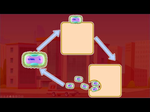
,1,In this visual lecture Dr. Aizaz talks about Replicative cycle of poxviruses and Tecovirimat
Life cycle is the series of changes that the organism goes through after it enters the cell and then it forms a new generation of virus which in turn infect more cells. Since the viruses aren’t considered a true living thing because they cannot survive outside the host, the term replicative cycle is used rather than “life cycle”
The replicative cycles of both monkeypox, as well as the smallpox virus, are based on studies done on vaccinia virus which is a virus belonging to same genus. It is presumed that smallpox and monkeypox viruses follow the same pattern of replicative cycle.
There are two types of poxviruses particles.
One is covered with the double envelopes is called extracellular virion. The other type is covered by a single envelope and is called mature virion.
When the extracellular virion’s outer membrane, via its tubules, interacts with the GAGs [need more info on GAGs] of the cell [which cell], the outer membrane is spontaneously disrupted, such that the inner membrane (inner envelope) is exposed. This inner membrane than fused with cell membrane and the nucleocapsid core along with lateral bodies, is ferried inside the cell.
The mature virion has a single membrane which can directly fuse with the cell membrane and the nucleocapsid core is, in turn, transported into the cell.
The mature virion can also be endocytosed inside the cell. It binds with a particular receptor [GAGs] on the cell membrane and the part of membrane pinches inward to form vesicle containing virion. It later sort of hatches out into the cytoplasm.
The virus cannot do much without expressing its genome, the DNA. So it must expose the DNA. The problem is that it is coated the core. To uncoat it needs certain proteins.
The virus brings its own DNA dependent RNA polymerase that forms mRNA for some early proteins including those that are required for uncoating. These mRNA transcripts are very small and are sent out of the capsid core into the cytoplasm. Here, it uses the host’s cytoplasmic ribosomes to form proteins. A few of the early proteins fully uncoat the viral DNA.
The viral DNA also contain gene for DNA polymerase / replicase, that creates a number of copies of the DNA to packed in the next generation virus particles.
The structural proteins like core proteins and the proteins of lateral bodies are also synthesized within the cytoplasm and they bind with the one of the copies of DNA to assemble an immature virus particle, that isn’t yet enveloped. Other proteins like RNA polymerase are also packed within the nucleocapsid core.
Some of the viral proteins are studded into Endoplasmic reticulum and these portions of ER buds off and surround the immature virion to form the mature virion. Some of the mature virions are wrapped by another membrane from the Golgi apparatus. This virion is then exocytosed out of the cell as an extracellular virion. The mature virion is released as a result of cell death when cells are broken (cell lysis). These released virions are now ready to infect other cells and as they do so the cycle continues.
The entire replicative cycle of poxvirus occurs within the cytoplasm
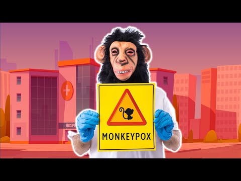
,1,In the beginning of May 2022, a British resident presented to the hospital in UK with Smallpox/Monkeypox like symptoms. He had history of travel to Nigeria, where the Monkeypox is endemic (regularly found among particular people or in a certain area).
Diagnosis of monkeypox was established by the Rare and imported pathogens laboratory of UK via the PCR. This marks as an index case of Monkeypox. After mid of May onwards, several cases of Monkeypox were reported in different countries across the world. In accordance with the niche of this channel, I won’t be telling you the number of cases, nor will I go into the news aspect of this outbreak. My aim is to educate you about the scientific aspects of Monkeypox virus and this disease.
Both smallpox and monkeypox viruses belong to a family of viruses called poxviridae; named so because they form the pockmarks (Pitted scar marks on the skin).
The poxviruses cause disease in vertebrates and insects. The subfamily that causes disease in vertebrates is called chordopoxviruses. It is further divided in `1to several genera. One of them is Orthopoxviruses. Some of the important species of this genus are monkeypox, smallpox, cowpox, vaccinia virus etc. All these are very similar structurally as well as clinically. It’s hard to differentiate between them without genetic testing like PCR.
The smallpox virus has no animal reservoir. However, monkeypox has animal reservoirs like squirrels and other rodents. Actually, monkeypox is a misnomer. Monkeys are not major reservoirs of this virus. However, it is named so because it was first found in laboratory monkeys in 1958.
Monkeypox is not a new disease nor is the Monkeypox virus a new virus, unlike COVID-19. Monkeypox virus has been known for decades. It is closely related to the smallpox virus which was eradicated in 1980. Monkeypox is endemic in some African countries. Monkeypox disease is almost indistinguishable clinically from smallpox disease, but it is typically less fatal and less contagious than smallpox.
But are we suddenly concerned about Monkeypox virus?
Historically, monkeypox has been mainly transmitted to humans via the animal reservoir through handling of animals and their meat. Human to human transmission has not been very common. But now we are seeing a surge in cases of people who do not have travel history to African countries or the history of handling animals or animal meat. This is concerning because there is a possibility that the virus might have gotten a mutation that may have increased its ability of human-to-human transmission. Its DNA virus. Mutations are quite rare as compared to RNA viruses like coronavirus, but still it’s a possibility.
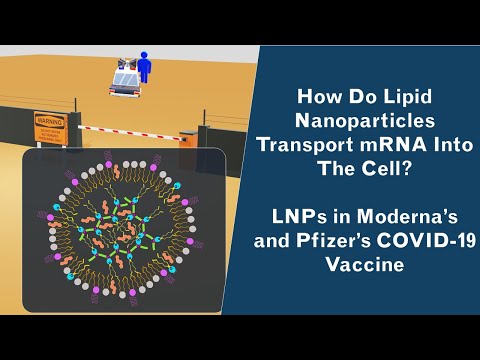
,1,In this video, Dr. Aizaz from Medicovisual describes how Lipid Nanoparticles work and what is their structure.
Previously we have already discussed how mRNA vaccines work. The idea is to transport the mRNA of the spike protein of coronavirus into the cell. The cell will then create spike proteins and will train the immune system to recognize the Sars-CoV-2 from its spike protein.
The problem here is that a naked RNA cannot enter into the cell. And of course, without entry into the cell, the mRNA vaccine will never work. Furthermore, the RNA particles present outside the cell will be quickly degraded by the RNAses present in the extracellular environment.
To solve this problem, Lipid nanoparticles were created. These are incredibly small particles, measured at a nanoscale. [From your secondary school knowledge, you must be knowing that 1 nm = 10-9m]. These tiny lipid nanoparticles are made up of fat/lipids. They can keep the RNA safe from the RNAses and can faithfully transport the RNA inside the cell, into its cytosol.
But the question is why does mRNA, which is a type of RNA by the way, cannot, by itself, cross the cell membrane and enter into the cell? Why does it need external help from either a viral vector or a lipid nanoparticle?
The reason is that RNA [and DNA too] is negatively charged, because of the presence of negatively charged Phosphate ions attached with each of its structural subunits; nucleotides. So, it is the Phosphate ions that impart a net negative charge to the RNA and DNA.
Charged or ionic substances have a hard time crossing the cell membrane made up of lipid bilayer.
When surrounded by the lipid nanoparticle, RNA can cross the lipid-bilayered cell membrane as it becomes a lipophilic substance.
You can imagine it from a simple analogy. Let us suppose you want to a restricted area where only Police is allowed to go. Of course, you will be denied entry at the gate. [Think of the gate as the cell membrane].
You still want to go to that restricted area. You call your friend who is a police officer. You sit in his police van and then easily pass through the security checkpoint.
The same is happening here. Foreign RNA is not allowed to go inside, so it enters into this police van or LNP to cross the cell membrane.
After understanding why there is a need for a lipid nanoparticle. Let us now discuss what is it made up of and then will understand how it works.
Structure and components of LNPs
As discussed earlier, RNA is negatively charged. You must be aware of the fact that opposite charges attract. So, cationic lipids [positively charged ions] will attract and thus bind with the negatively charged [Anionic] RNA.
Cationic lipids contain an amine group head that has a positive charge on it. Along with that, it has legs made up of fatty acid chains, that are non-polar or non-ionic.
When such cationic lipids come across the RNA, they will obviously orient their charged [cationic] amine head towards the RNA and will orient the legs outward.
Permanently cationic [having a permanent positive charge] lipids are toxic and can disrupt the integrity of cell membrane [1]
To mitigate this problem, ionizable cationic lipids are used. These lipids are either neutral or slightly charged [cationic] at physiological pH. But in the lab, they create the vaccine at an acidic pH.[2] On acidic pH, the Proton or H+ ion quickly attaches with the amine head of the lipid causing it to acquire a positive charge. Due to this positive charge, it embraces and encapsulates the negatively charged RNA.
At physiological pH, as these ionizeable cationic lipids will become neutral, they may cease to embrace the RNA. So this particle is further surrounded by one or more additional layers of phospholipids. Thus RNA will still remain packed and protected inside the core of lipid nanoparticle.
They are mostly non-cationic structural lipids with some cationic lipids too embedded in between them, here and there.
Then there are some lipids, conjugated with a special molecule called PEG [Polyethylene glycol], that too are embedded into this outer layer. The outer layer of PEG prevents the aggregation or fusion of LNP particles with each other due to its steric hindrance [3]
Polyethylene glycol offers a steric hindrance and prevents the immediate take-up of these lipid nanoparticles by immune cells followed by their destruction. [3] Thus, it slows down the clearance of these LNPs quickly from the body.
PEGylation is reversible in such a way that LNPs gradually undergo de-PEGylation. PEG is gradually removed and the de-PEGylated LNP is then picked up by the cells gradually.
Cholesterol is also present interspersed in the spaces between the nanoparticles. It maintains the integrity and imparts the structure stability to LNP. Cholesterol, and structural phospholipids, forming the outer layer of LNP, are also called helper lipids
Website: https://www.medicovisual.com
Email: draizaz@medicovisual.com